



Dental implants can replace missing teeth, but they only work well if your jawbone is healthy and strong enough to support them.
Before placing an implant, dentists need to check if you have enough bone and if the bone quality is good.
Dentists assess bone quality before implant surgery using clinical exams, X-rays, and advanced 3D imaging like CBCT scans to measure bone density, volume, and structure.
The evaluation of your jawbone and gum tissue helps your dentist create a safe treatment plan. Good bone quality for dental implants means your implant has a better chance of lasting for many years.
Poor bone quality might mean you need extra treatment before getting an implant.
Understanding how dentists assess bone density can help you feel more prepared for your consultation.
This article explains the different methods dentists use to check your bone and what happens if your bone needs improvement before implant placement.
Key Takeaways
- Dentists use clinical exams and imaging tools to measure your bone quality and volume before placing implants
- CBCT scans provide detailed 3D images that show bone density and help plan the exact implant position
- Bone grafting may be needed if you don’t have enough healthy bone to support an implant
The Importance of Bone Quality in Dental Implants
The quality of your jawbone determines whether an implant can anchor properly and last for years.
Your bone’s structure and density directly affect how well the implant fuses with surrounding tissue and stays stable under daily chewing forces.
Role in Implant Stability and Longevity
Your jawbone needs to be strong enough to hold an implant in place from the moment it’s inserted. When you have good bone quality, the implant stays firmly positioned during the healing process and beyond.
Weak or soft bone can cause the implant to move or fail over time.
The amount of stress your implant can handle depends on your bone’s ability to support it. When you chew food, your implant experiences significant force that transfers directly to the surrounding bone.
Dense, healthy bone absorbs these forces better than poor-quality bone.
Bone quality affects implant success because it determines how well your jawbone can maintain the implant’s position over many years.
If your bone lacks sufficient strength or volume, your dentist may recommend bone grafting before placing the implant.
Impact on Osseointegration
Osseointegration is the process where your bone cells grow directly onto the implant’s surface, creating a permanent bond. This biological connection takes several months to develop fully.
Your bone quality plays a major role in how quickly and completely this fusion occurs.
Higher-quality bone with good mineral content supports faster osseointegration. Your bone cells need adequate density and structure to attach properly to the titanium implant surface.
Poor bone quality can slow down this process or prevent complete integration.
The success of your dental implant depends on proper osseointegration. Without this biological bond, your implant won’t function like a natural tooth root and may fail prematurely.
Bone Quality Versus Bone Density
Bone quality and bone density are related but different concepts. Your bone density refers to how much mineral content exists in a specific volume of bone.
Bone quality includes density plus other factors like bone structure, thickness, and overall health.
Dentists often consider bone quality equivalent to bone density when viewing X-rays, but quality involves more than just how dense your bone appears. Your bone’s architecture and ability to remodel itself also matter for implant success.
You might have adequate bone density but poor bone quality if the bone structure is compromised. Medical conditions, medications, or previous tooth loss can affect your bone quality even when density measurements seem normal.
Clinical and Radiographic Methods for Bone Assessment
Dentists use both hands-on examination and imaging tools to evaluate the bone where your implant will be placed. These methods help determine bone density, bone quality, and the position of important anatomical structures.
Clinical Examination and Palpation
Your dentist starts by examining your mouth directly with their hands and instruments. They press on your gums to feel the underlying bone and check its thickness and firmness.
This hands-on approach gives them a sense of how much bone is present.
During palpation, your dentist can identify areas where bone may be too thin or soft. They also look at your gum tissue health and the space between teeth.
Clinical examination helps spot obvious problems before any imaging happens.
However, this method has limits. Your dentist cannot see through your gums to know exactly what’s underneath.
They cannot measure precise bone dimensions or see internal bone structure with their hands alone. That’s why diagnostic imaging becomes necessary for a complete assessment.
Periapical and Panoramic Radiographs
A periapical radiograph shows a detailed view of one or two teeth and the bone around their roots. Your dentist uses these X-rays to check bone height and look for infections or bone loss.
These images provide good detail for small areas.
A panoramic radiograph captures your entire jaw in one image. This X-ray shows all your teeth, both jaws, and surrounding anatomical structures like sinuses and nerves.
Traditional radiographic evaluation helps dentists plan where implants might fit.
Both types give your dentist a two-dimensional view of your bone. They can measure vertical bone height and estimate bone density by looking at how light or dark the bone appears on the film.
Limitations of Traditional Radiography

Two-dimensional X-rays cannot show bone width or depth accurately. You need three-dimensional information to know if enough bone exists in all directions for an implant.
Flat images also make it hard to see exactly where nerves and blood vessels are located.
These traditional X-rays can miss important details about bone quality. The bone might look adequate on a flat image but actually be too thin when viewed from another angle.
Overlapping structures can hide problems or make bone appear thicker than it really is.
Measuring bone density from these images relies heavily on your dentist’s judgment. Different dentists might interpret the same X-ray differently, which can affect treatment planning.
Advanced Imaging: The Role of CT and CBCT
Three-dimensional imaging technologies provide detailed views of your jawbone structure that traditional x-rays cannot capture.
These advanced tools measure bone density, volume, and proximity to vital anatomical structures with remarkable precision.
Computed Tomography for Bone Evaluation
Computed tomography scans create cross-sectional images of your jaw that reveal bone quality in fine detail.
Your dentist can measure bone density values directly from CT images, which helps determine if your bone is strong enough to support an implant.
CT scans offer excellent contrast resolution. This means they can distinguish between different tissue types more clearly than some other imaging methods.
The technology is particularly useful when your dentist needs to evaluate soft tissue alongside bone structures.
Medical CT scanners provide highly accurate measurements. However, they typically deliver higher radiation doses compared to dental-specific imaging options.
Modern multi-slice CT machines have improved significantly, with some newer models achieving radiation levels comparable to other dental imaging techniques.
Cone Beam CT (CBCT) in Dental Implant Planning
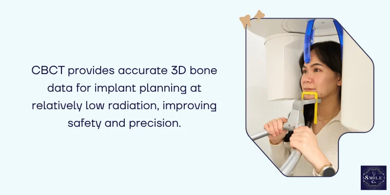
Cone beam computed tomography has become essential in implant dentistry because it provides three-dimensional jaw bone data at reasonable costs and radiation doses.
CBCT scanners are compact enough to fit in dental offices, giving you convenient access to advanced imaging.
Your CBCT scan reveals bone height, width, and density at the proposed implant site. The technology offers voxel sizes between 0.08 and 0.4 mm, providing the spatial resolution needed to visualize small anatomical details.
Radiation doses from CBCT vary widely between machines, ranging from the equivalent of 2 to 200 panoramic radiographs. Your dentist should use optimized protocols that balance image quality with radiation exposure.
Segmentation accuracy can reach 200 micrometers on quality systems, though some machines may have larger inaccuracies.
3D Imaging for Anatomical Structure Assessment
Three-dimensional scans map critical structures near your implant site, including nerves, blood vessels, and sinus cavities.
CBCT provides a detailed evaluation of anatomical structures, offering precise information on bone quality and the location of vital areas that must be avoided during surgery.
Your dentist examines the inferior alveolar nerve in the lower jaw and the maxillary sinus in the upper jaw. These structures require careful measurement to maintain safe distances during implant placement.
The mandibular canal housing the inferior alveolar nerve needs at least 2mm of clearance from the implant.
Digital planning software allows your dentist to measure exact distances and angles from the 3D scan. This reduces risks like nerve injury or sinus perforation during the procedure.
Bone Grafting and Site Enhancement Strategies
When your jawbone lacks adequate volume or density for implant placement, dentists use bone grafting techniques to create a stable foundation that supports successful osseointegration.
Indications for Bone Grafting
You may need bone grafting if you’ve experienced significant bone loss from tooth extraction, periodontal disease, or trauma.
Bone augmentation helps provide sufficient quality and quantity of bone in areas where the ridge has deteriorated.
Your dentist will recommend grafting when measurements show inadequate bone width (less than 6mm) or height for standard implant placement.
Patients who have worn dentures for extended periods often require grafting because the jawbone gradually resorbs without tooth roots to stimulate it.
Long-term tooth loss creates atrophic ridges that cannot accommodate implants without enhancement.
Your bone quality also matters, if imaging reveals low-density bone that won’t support initial implant stability, grafting can strengthen the site before surgery.
Techniques to Improve Bone Quality and Density
Block grafts involve harvesting bone from another area of your mouth and securing it to the deficient site. This technique achieved survival rates ranging from 97.3% to 100% across multiple studies, with a mean of 98.5%.
Particulate grafts use smaller bone particles to fill defects and build volume. Your dentist may use autologous bone (from your own body), allografts (donor bone), xenografts (animal-derived bone), or synthetic materials.
Blood derivatives and composite bone grafts combine multiple materials to enhance healing. Blood derivatives showed 94.7% survival rates, while composite grafts achieved 99.3% survival rates but lower success rates at 90.7%.
Ridge augmentation techniques add height or width to your jawbone. Guided bone regeneration uses membranes to protect the graft while new bone forms over 4-6 months.
Outcomes and Predictability of Grafting
Autologous bone grafts typically provide the most predictable results because your body readily accepts its own tissue.
These grafts demonstrated higher success rates and fewer complications compared to other materials in long-term studies.
The healing period after grafting usually takes 4-6 months before implant placement. During this time, the graft material integrates with your existing bone to create sufficient bone density for implant support.
Success rates vary by graft type and location. Studies with 3-5 year follow-ups show that properly placed grafts remain stable and support functioning implants.
Your dentist monitors graft integration through periodic imaging to confirm adequate bone quality has developed before proceeding with implant surgery.
Integrating Diagnostic Findings Into Implant Treatment Planning
After gathering diagnostic information, dentists use these findings to create a precise plan for your implant surgery.
This process involves checking if you’re a good candidate, identifying the exact placement location, and adjusting the approach based on your unique bone structure and anatomy.
Evaluating Implant Suitability
Your dentist reviews all diagnostic data to determine if dental implants will work for you. They look at bone density measurements, bone mineral content, and overall bone volume at the proposed implant site.
If your bone quality falls below certain standards, your dentist might recommend bone grafting procedures before placing the implant.
Diagnostic treatment planning is crucial to achieving optimal success in oral rehabilitation. Your general health also plays a role in this assessment.
Conditions like uncontrolled diabetes or osteoporosis may affect bone healing and implant success rates.
The evaluation includes measuring the height and width of available bone. Most implants need at least 10mm of bone height and 6mm of width for stable placement.
Mapping Implant Location Relative to Anatomical Structures
Your dentist uses diagnostic imaging to map out vital structures near the implant site. The mandibular canal in your lower jaw contains important nerves and blood vessels that must be avoided. In your upper jaw, the maxillary sinus sits above the back teeth area.
Placement of implants in sites with inadequate bone may cause complications such as impingement on these vital anatomical structures.
Your dentist measures the exact distance from the planned implant position to these areas. They typically maintain a safety margin of at least 2mm from nerves and sinuses.
Digital planning software allows your dentist to visualize the implant position in three dimensions. This helps ensure the implant angle and depth won’t damage surrounding teeth roots or nerves.
Personalizing Treatment Based on Imaging Results
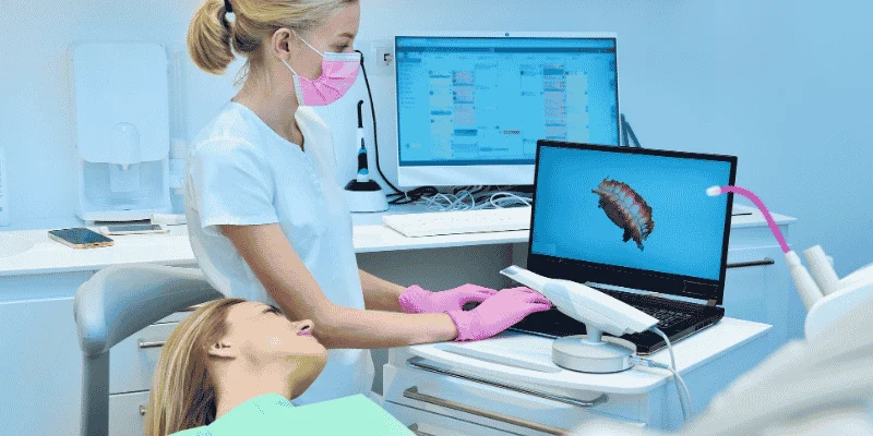
Your implant planning becomes tailored to your specific anatomy and bone characteristics. If imaging shows thinner bone on one side, your dentist might choose a narrower implant or adjust the placement angle.
Dense bone may require special drilling techniques to prevent overheating during surgery.
Some patients need surgical guides created from their imaging data. These guides fit over your teeth or gums during surgery and direct the drill to the exact planned position.
Your dentist also selects the appropriate implant length and diameter based on your available bone.
Common personalized adjustments include:
- Modified implant sizes (diameter and length)
- Adjusted placement angles
- Staged procedures with bone grafting
- Custom surgical guides
- Alternative implant designs for specific bone types
Frequently Asked Questions
Dentists use specific imaging tools and measurement systems to determine if your jawbone can support an implant.
Understanding bone density classifications and minimum dimension requirements helps explain when additional procedures might be necessary.
What imaging tests will my dentist use to check if I have enough jawbone for a dental implant?
Your dentist will typically use cone beam computed tomography (CBCT) scans to evaluate your jawbone before implant surgery.
CBCT scanning is the most common method for measuring bone density and examining the three-dimensional structure of your jaw.
This type of scan shows your dentist the exact height, width, and depth of bone available at the implant site. It also reveals important details about bone quality that regular X-rays cannot show.
Some dentists may also use panoramic X-rays as an initial screening tool. These give a broad overview of your entire jaw but provide less detail than CBCT scans.
How does my dentist decide whether my bone height and width meet the minimum requirements for an implant?
Your dentist measures the bone dimensions at the proposed implant site using the CBCT scan images. Most dental implants need at least 10-12 millimeters of bone height and 5-6 millimeters of bone width to be placed safely.
The measurements help your dentist select the right implant size for your specific situation. If your bone falls short of these minimum dimensions, you may need bone grafting before implant placement.
Bone quality assessment looks at density, width, height, and architecture to create a complete picture of your jawbone condition.
Your dentist also checks the distance between the implant site and important structures like nerves and sinuses.
What do the D1, D2, D3, and D4 bone density types mean, and why do they matter for implant success?
The D1 through D4 classification system describes how dense your jawbone is. D1 bone is the densest and hardest, made mostly of compact bone with very little soft inner bone.
D2 bone has a thick outer layer of compact bone surrounding porous inner bone. This density type offers excellent support for implants and is considered ideal for implant placement.
D3 bone features a thin outer layer with porous inner bone of moderate density. D4 bone is the least dense, with a very thin outer shell and soft inner bone that provides the weakest support.
Bone density directly affects implant stability and how well the implant integrates with your jawbone. Denser bone types generally lead to better initial stability and higher success rates.
In which parts of the mouth are different bone density types most commonly found?
The front part of your lower jaw typically has the densest bone, usually D1 or D2 type. This area provides the strongest foundation for dental implants.
The back of your upper jaw tends to have the softest bone, often D3 or D4 type. This makes implant placement in this area more challenging and may require special techniques.
The front of your upper jaw and the back of your lower jaw usually fall somewhere in between, with D2 or D3 bone density. Your dentist considers these natural variations when planning your implant treatment.
When would my dentist recommend a bone graft before placing an implant?
Your dentist may recommend bone grafting if you don’t have enough bone height or width at the implant site. This procedure adds bone material to build up the area before implant placement.
Bone grafting is also needed when your bone density is too low to support an implant. After grafting, the bone must integrate and heal before implants can be placed, which extends your treatment timeline but improves long-term success.
If you’ve had missing teeth for a long time, your jawbone may have shrunk significantly. You might also need grafting if you’ve experienced bone loss from gum disease or tooth infection.
What is the “50-40-30 rule” in implant planning, and how does it relate to bone support?
The 50-40-30 rule guides dentists in planning how much bone should surround your implant. This rule suggests that at least 50% of the implant length should be anchored in bone for adequate support.
The numbers also indicate that 40% bone-to-implant contact is acceptable in some cases, while 30% represents the minimum threshold for implant stability.
Following this rule helps ensure your implant has enough bone support to function properly.
Your dentist uses these guidelines along with measurements of bone height and width to determine if your jawbone can support an implant.
If your bone doesn’t meet these criteria, grafting or alternative treatment options may be necessary.